
Talking through the diagnosis, step by step.
A swollen abdomen, fluid inside the abdomen and a raised CA-125 blood test can make doctors worry about ovarian cancer. These findings do not always mean cancer. In India, abdominal tuberculosis (TB) can look remarkably similar, and the two conditions need very different treatment. Getting the diagnosis right before major surgery or chemotherapy is one of the most important steps in care.
What to Do If Ascites and an Ovarian Mass Are Found
Ascites (fluid in the abdomen) together with an ovarian or pelvic mass needs specialist evaluation. Neither finding proves cancer, and neither proves TB. Ask for a gynaecologic oncology opinion, a good-quality ultrasound, and a clear plan for how the diagnosis will be confirmed before any major treatment begins.
The rest of this article explains each step. For symptoms that should prompt a doctor's visit, see our article on ovarian cancer symptoms.
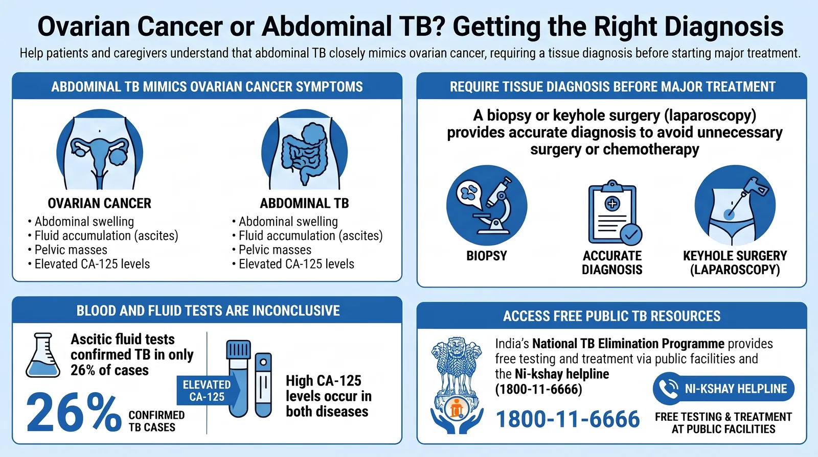
Ovarian cancer and abdominal TB can look alike. A tissue diagnosis guides the right treatment.
Why Can Ovarian Cancer and Abdominal TB Look So Similar?
Both conditions can cause abdominal swelling, pain, loss of appetite, weight loss, ascites, thickening of the peritoneum (the membrane lining the abdomen) and lumps in the abdomen or pelvis. Both can also raise CA-125.
A 2020 study from Christian Medical College, Vellore, reviewed 23 women who had been referred for suspected ovarian or peritoneal cancer and were ultimately diagnosed with abdominal TB. Their mean CA-125 was 333.5 U/mL. The authors concluded that abdominal TB is common in India, can mimic ovarian cancer, and needs a high index of suspicion.[1] This was a retrospective study of a selected group, so it shows how TB can mislead, not how often it does.
"This article does not suggest that doctors should assume TB rather than cancer. Both are serious, and both must be considered."
The aim is a reliable diagnosis before a patient has major surgery or chemotherapy for the wrong condition, or waits too long for the right one.
What Is Ascites, and Does It Always Mean Cancer?
No. Ascites is an abnormal build-up of fluid in the abdominal cavity. It is a sign that something needs investigating, not a diagnosis in itself.
People with ascites may notice a growing or persistently bloated abdomen, heaviness, feeling full after a small meal, or breathlessness when fluid presses against the diaphragm.
Many conditions cause ascites. These include ovarian, tubal or peritoneal cancer, other abdominal cancers, peritoneal TB, chronic liver disease, heart failure and some less common inflammatory conditions.
The UK's National Institute for Health and Care Excellence (NICE) advises a specialist referral for women with ascites or a pelvic or abdominal mass on examination, unless the mass is clearly uterine fibroids.[2] NICE is a UK body, and its recommendations are not automatically the standard in India. The principle, however, is that ascites needs proper investigation.
Can Ovarian Cancer Be Mistaken for TB?
Yes. Published case series from several countries show how easily this happens. In some cases, women were first taken to surgery for suspected cancer and the final diagnosis was TB.
- India (2020): In the Vellore series, ascitic fluid confirmed TB in 26% of women. Omental biopsy, which samples the fatty tissue in the abdomen, showed TB in 69%. The authors suggested that diagnostic laparoscopy, a keyhole procedure that lets the surgeon see inside the abdomen and take targeted samples, could be considered early in the work-up rather than after repeated inconclusive tests.[1]
- Ethiopia: Four women were each first thought to have ovarian cancer and were taken to surgery. The surgeon found typical features of TB and took a biopsy, which confirmed pelvic TB. The authors noted that radical operations such as hysterectomy and removal of the ovaries and tubes could have harmed these women's health and fertility.[3]
- Mexico (2022): Nine women with abdominal TB that resembled advanced ovarian cancer were reviewed. None had a history of lung TB. Even during surgery, 62.5% were first diagnosed as cancer on intraoperative pathology. The authors described TB as "the great imitator."[4]
- Nigeria (2026): A retrospective series of three women, aged 18, 44 and 56, had ascites, adnexal masses, peritoneal thickening and raised CA-125. Surgical specimens showed granulomatous inflammation consistent with TB in all three. The authors recommended a high index of suspicion and early minimally invasive tissue diagnosis in TB-endemic settings.[5]
These reports illustrate diagnostic difficulty. They do not show how often misdiagnosis happens in India or elsewhere.
Can a Doctor Tell the Two Apart From Symptoms Alone?
Usually not with certainty. The symptoms overlap, and many people with abdominal TB have no cough, fever or known history of TB. The absence of these features does not rule TB out. A history of TB does not rule cancer out either.
Symptoms guide which tests are needed. They do not replace the tests.
Is Ultrasound Enough to Diagnose Ovarian Cancer?
No. Ultrasound is the usual first imaging test for an ovarian or pelvic mass, but it cannot confirm or exclude cancer on its own.
Ultrasound shows whether a lump is solid, fluid-filled or mixed, whether it has thick walls or solid areas, whether blood flow is abnormal and whether there is ascites. The European consensus statement on preoperative diagnosis of ovarian tumours, from ESGO, ISUOG, IOTA and ESGE, found that expert ultrasound assessment and validated ultrasound-based models, such as IOTA models, are particularly useful for distinguishing benign from malignant masses.[6] The quality of the scan and the experience of the person reading it matter a great deal.
Transvaginal ultrasound gives more detail of the ovaries and pelvic organs. Transabdominal ultrasound gives a wider view and is useful for large masses or heavy ascites. The two are complementary.
Specialists use a standard reporting system called O-RADS (Ovarian-Adnexal Reporting and Data System). It describes ovarian lumps and estimates cancer risk from their appearance, on a scale from O-RADS 0 (incomplete assessment) to O-RADS 5 (high risk).[7] It supports clinical judgement and does not replace tissue diagnosis.
A reassuring scan does not rule out cancer if symptoms persist.
What Does CA-125 Tell Us, and What Does It Not?
CA-125 is a blood protein used in assessing and monitoring many ovarian cancers. It is useful, but it is not specific to cancer. Many laboratories use 35 U/mL as the usual upper reference limit. That figure is a laboratory reference point, not a universal diagnostic threshold.
NICE's guidance shows why age matters:[2]
- Women aged 40 to 49: 35 IU/mL is used as the threshold for arranging an ultrasound in women with persistent symptoms.
- Women over 50: NICE uses age-specific thresholds in 10-year bands, which are lower than 35 IU/mL in several age groups.
- Women 39 and under: NICE says CA-125 is not accurate enough to guide decisions, because ovarian cancer is rare in this age group and a normal result can give false reassurance.
In every age group, NICE advises that CA-125 should not be used alone to make decisions about suspected ovarian cancer. Check the current NICE guidance before relying on specific numbers, as thresholds are reviewed from time to time.
Practical points:
- A high CA-125 does not prove cancer. It can rise with endometriosis, menstruation, pregnancy, pelvic infection, liver disease and peritoneal inflammation, including TB.
- A normal CA-125 does not rule out cancer. Early-stage cancers and some subtypes may not raise it.
- Peritoneal TB can raise CA-125 substantially. A high result should prompt careful investigation, not an automatic cancer or TB label.
What Are HE4 and ROMA?
HE4 is another blood protein that may be raised in some ovarian cancers. ROMA (Risk of Ovarian Malignancy Algorithm) combines CA-125, HE4 and menopausal status to estimate the chance of cancer in a woman with a pelvic mass.
The ESGO, ISUOG, IOTA and ESGE consensus addresses these biomarkers and prediction models, but places expert ultrasound assessment at the centre of pre-operative evaluation.[6] HE4 and ROMA are not needed for every patient, and they do not replace imaging or tissue diagnosis. Their results depend on the assay and laboratory used, so they should be interpreted with a doctor.
Is a CT Scan Needed?
Often, yes. When malignancy is suspected, CT of the abdomen and pelvis, with chest imaging when indicated, is central to assessing how far disease extends. It helps show peritoneal and omental involvement, enlarged lymph nodes and any spread to other organs. This information guides surgical and treatment planning.
CT cannot reliably distinguish cancer from abdominal TB on its own. Both can cause ascites, peritoneal thickening and nodules. CT findings can strengthen or weaken suspicion, but tissue sampling may still be needed.
Is PET-CT Needed?
Not routinely. PET-CT is used for particular clinical questions. It measures metabolic activity, and inflammation from infection, including TB, can also appear active, so it cannot reliably separate cancer from infection in every case. For most initial assessments of a suspected ovarian mass, ultrasound, appropriate CT and tissue diagnosis are more central. Ask your doctor whether a test is likely to change the treatment plan.
What Is FNAC, and Can It Tell TB From Cancer?
FNAC stands for fine-needle aspiration cytology. A thin needle collects cells, which are examined under a microscope. It can be useful for sampling an accessible lesion or enlarged lymph node, and image-guided sampling is one of several options for obtaining tissue.
FNAC has limits:
- It may not collect enough cells.
- The needle may miss the part of the lesion that matters.
- Cytology may not show enough tissue structure to classify a tumour fully.
- The term is sometimes used loosely. Sampling fluid is not the same as taking a core biopsy from an abdominal deposit or a lymph node.
Granulomas, which are clusters of immune cells, can support TB, but they are not exclusive to TB. Malignant cells establish cancer in the sampled material, though further tests may be needed to identify the primary site. A negative or inconclusive FNAC does not exclude either disease.
What Do Ascitic Fluid Tests Show?
If there is significant ascites, a doctor may drain a sample, which is called paracentesis. The fluid can be checked for malignant cells, white blood cells, protein and albumin. It can also be tested for TB bacteria, using microscopy, culture or molecular tests such as GeneXpert, and sometimes for adenosine deaminase (ADA), a marker that can support TB in the right clinical setting.
Fluid tests have limits:
- Malignant cells may be absent even when cancer is present.
- TB bacteria are often very few in abdominal TB, so smears, cultures and molecular tests can all be negative.
- In the Vellore series, ascitic fluid confirmed TB in only 26% of women who were ultimately diagnosed with it.[1]
A negative ascitic fluid result does not exclude abdominal TB. Likewise, it does not exclude ovarian cancer. When fluid tests are inconclusive and suspicion remains, tissue sampling may be needed.
How Is Abdominal TB Confirmed?
Diagnosis usually combines several strands of evidence. These may include blood tests, a chest X-ray to look for TB in the lungs, ascitic fluid analysis (including TB tests where available), and tissue sampling when fluid results are inconclusive.
Tissue can be obtained in more than one way:
- Image-guided biopsy of an accessible peritoneal or omental lesion, or an enlarged lymph node.
- Diagnostic laparoscopy, a keyhole procedure that allows direct inspection and targeted biopsy.
Which route is right depends on where the disease is, what can be safely reached, what is available locally and what the treating team is trying to confirm. Diagnostic laparoscopy is not automatically the first step for everyone. In a selected patient who has inconclusive results and remains a diagnostic dilemma, earlier tissue diagnosis may avoid repeated tests and unnecessary delay.
The Indian national guidelines for extrapulmonary TB, INDEX-TB, describe both fluid-based and tissue-based approaches. They should be applied to each patient rather than followed as a fixed checklist.[8]
Why Is Tissue Diagnosis So Important Before Treatment?
Because the two conditions need very different treatment. Ovarian cancer is usually treated with surgery and chemotherapy. Abdominal TB is usually treated with a standard multidrug anti-TB regimen. Starting the wrong treatment can delay the right one and expose a patient to avoidable harm.
The regimen and duration of TB treatment are determined by the treating team under current TB guidance, and six months is often used for drug-sensitive disease.
ASCO's resource-stratified guideline for ovarian masses states that ovarian cancer is diagnosed by histologic confirmation in all settings. Where resources allow, CT-guided biopsy or laparoscopy is preferred to obtain tissue, and treatment requires histologic confirmation.[9] When advanced disease is suspected and chemotherapy is being considered before surgery, obtaining tissue first is the expected standard.
If surgery is the first step, the diagnosis may come from the surgical specimen. Whether a needle biopsy is needed beforehand depends on the lesion and the planned operation, and is a decision for the treating team.
India's ICMR published a consensus document on managing epithelial ovarian cancer in 2019. Its literature review runs to October 2015, so it should be read alongside more recent evidence.[10]
Can Ovarian Cancer Be Mistaken for a Benign Cyst?
Yes. Some cancers look relatively benign on imaging, and many complex ovarian cysts are harmless. Thick walls, solid areas, papillary projections and abnormal blood flow raise suspicion. A simple thin-walled cyst is usually less worrying, though follow-up depends on size, age, menopausal status and symptoms.
A normal CA-125 should not make a suspicious mass look reassuring. A complex cyst is not automatically a cancer.
Why Does This Matter in India?
Late presentation is a major challenge. A 2026 study from a tertiary cancer centre in Patna, Bihar, reviewed 437 women with confirmed ovarian cancer. Of these, 74.6% presented at FIGO stage III or IV, and over 75% came from rural backgrounds.[11] This was a single-centre retrospective study, so it does not represent national figures.
A hospital study from Tata Main Hospital, Jamshedpur, covering 2019 to 2024, found that 39.2% of women had stage III disease and 21.5% had stage IV at presentation.[12] This is also a single-centre study.
Taken together, these studies suggest that many women reach specialist care when disease is already advanced. That makes accurate early diagnosis more important.
Access to diagnosis is also a concern. Under the National TB Elimination Programme (NTEP), screening, diagnostic tests and treatment for TB, including extrapulmonary TB, are provided free at public health facilities.[13] The Ni-kshay Sampark helpline, 1800-11-6666, can direct patients to the nearest TB diagnostic and treatment centre.[14] Nutritional support is available for registered TB patients. For cancer care, patients should ask their doctor about public and cancer-centre options and whether the tests and treatment they need are available locally.
What Questions Should I Ask My Doctor?
- What do you think is causing the fluid or the lump, and what else could it be?
- Which tests will help tell cancer and TB apart?
- Is a tissue sample needed, and how will it be taken?
- Would a keyhole procedure (laparoscopy) help confirm the diagnosis sooner?
- Will any treatment start before we have a confirmed diagnosis?
- Should a gynaecologic oncologist review the case?
- Which tests are free in a public hospital, and which must be paid for?
- When should I seek medical care urgently?
When Should I Seek Medical Care?
Seek prompt medical review if you have:
- Abdominal swelling or ascites that is increasing or does not settle.
- Persistent bloating, abdominal pain, or feeling full very quickly.
- Unexplained weight loss or loss of appetite.
- A lump in the abdomen or pelvis, or an ovarian mass found on a scan.
- Fever, night sweats or a persistent cough, especially alongside these symptoms.
Go to the nearest emergency department without delay for severe abdominal pain, vomiting, difficulty breathing, fainting or a rapidly swelling abdomen.
The Key Message
Ascites, a raised CA-125, an ovarian mass or a negative fluid result cannot, on their own, establish a diagnosis. In India, where abdominal TB is an important alternative to ovarian cancer, the answer usually depends on careful clinical assessment, appropriate imaging and, when needed, tissue diagnosis obtained at the right time.
The aim is not to perform every available test. It is to gather enough reliable evidence to choose the correct treatment without avoidable delay or unnecessary surgery.
Frequently Asked Questions
Medical disclaimer: This article provides general educational information. It is not a substitute for individual medical assessment. Decisions about investigation and treatment should be made with your own doctor.
References
- Thomas A, Sebastian A, George R, et al. Abdominal tuberculosis mimicking ovarian cancer: a diagnostic dilemma. Journal of Obstetrics and Gynaecology of India. 2020;70(4):304–309. doi:10.1007/s13224-020-01322-8 link.springer.com
- National Institute for Health and Care Excellence (NICE). Ovarian cancer: recognition and initial management (CG122), quality statement on CA-125 age-specific thresholds (QS18). NICE. nice.org.uk
- Gashawbeza B, Tadessie U, Felagot T, Zelele D, Tesfaye HT. A case series of disseminated tuberculosis involving pelvic organs and peritoneum mimicking advanced ovarian malignancy. Journal of Gynecological Oncology, Vol. 3, Issue 3. remedypublications.com
- Morales-Castelán JE, Lara-Hernández ME, Hernández-Nava JM, Lino-Silva LS, Salcedo-Hernández RA. Abdominal tuberculosis mimicking ovarian cancer: a case series report. Gaceta Médica de México. 2022. researchexchange.ocrahope.org
- Offor JO, Achusi IB, Ajah LO. Abdominopelvic tuberculosis mimicking advanced ovarian malignancy: a case series emphasising diagnostic pitfalls in a tuberculosis-endemic setting. BMC Women's Health. 2026. link.springer.com
- Timmerman D, Fotopoulou C, et al. ESGO/ISUOG/IOTA/ESGE Consensus Statement on preoperative diagnosis of ovarian tumours. Ultrasound in Obstetrics & Gynecology / International Journal of Gynecological Cancer. 2021. pmc.ncbi.nlm.nih.gov
- American College of Radiology. O-RADS Ultrasound. ACR Clinical Resources. acr.org
- Central TB Division, Ministry of Health and Family Welfare, Government of India. INDEX-TB Guidelines: Guidelines on extra-pulmonary tuberculosis for India. Government of India. cochrane.org
- Vanderpuye VD, Clemenceau JRV, Temin S, et al. Assessment of adult women with ovarian masses and treatment of epithelial ovarian cancer: ASCO resource-stratified guideline. JCO Global Oncology. 2021. ace.asco.org
- Indian Council of Medical Research (ICMR). Consensus Document for Management of Epithelial Ovarian Cancer. 2019. icmr.gov.in
- Buddha Cancer Centre, Patna. A histopathological spectrum, descriptive epidemiology and survival status of ovarian cancer cases from a tertiary cancer centre in northern India: a retrospective cohort study. Malignancy Spectrum. 2026. eurekalert.org
- Tata Main Hospital, Jamshedpur. Epidemiological profile, clinical characteristics, and management of ovarian cancer in Eastern India: a hospital-based study (2019–2024 data). Cureus. doi:10.7759/cureus.101937 ncbi.nlm.nih.gov
- Ministry of Health and Family Welfare, Government of India. Rajya Sabha Unstarred Question No. 1733, answered 6 August 2024. Rajya Sabha. rsdebate.nic.in
- Press Information Bureau, Government of India. India Speeding Towards TB Elimination. 24 March 2026. pib.gov.in
Related Reading

Have an abdominal mass or fluid that needs evaluation?
Dr. Dipanwita Banerjee offers consultations at CNCI Kolkata for the evaluation of ovarian and pelvic masses and gynaecological cancers.