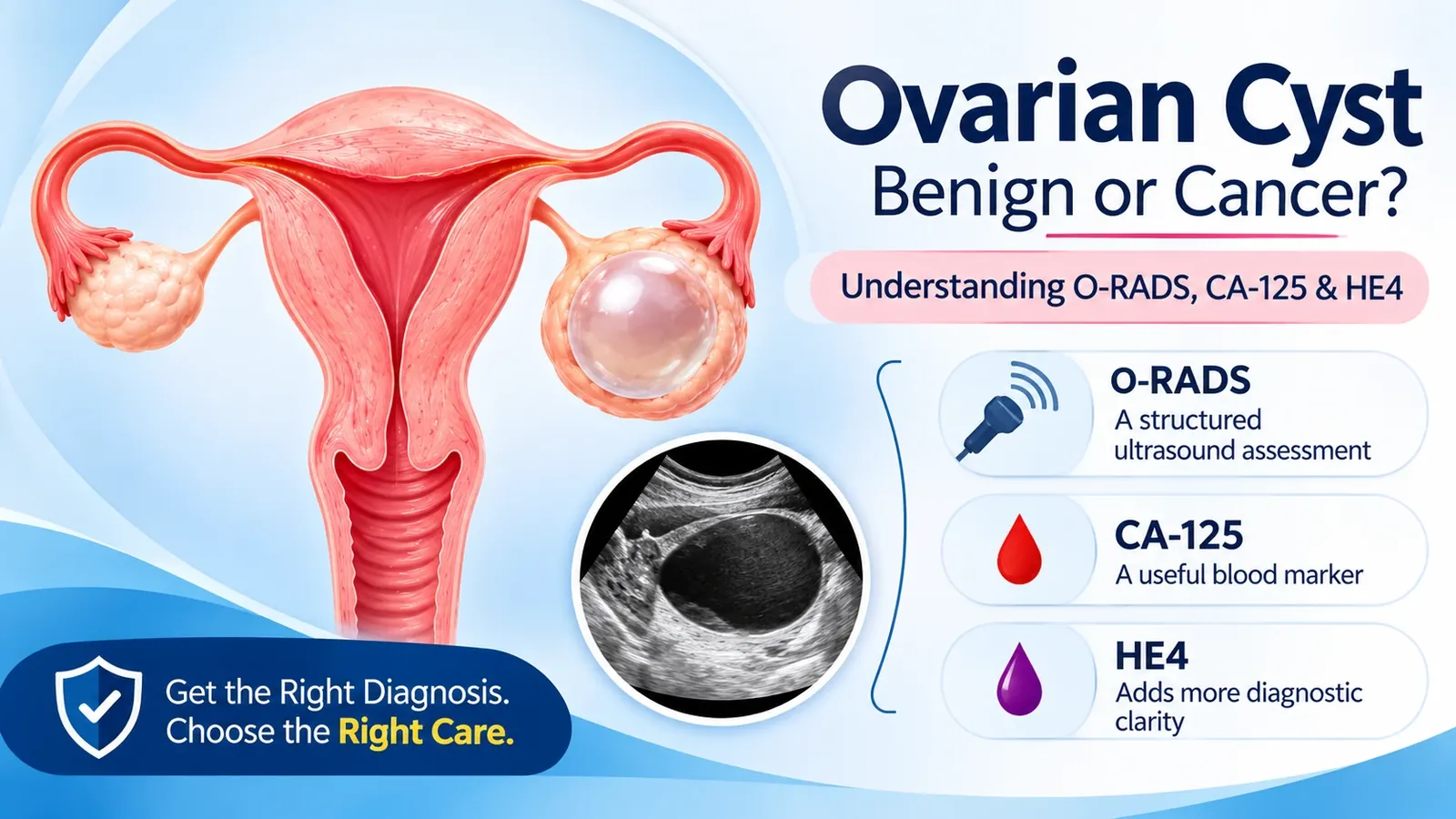
O-RADS, CA-125 and HE4: the tools used to assess an ovarian cyst.
Is every ovarian cyst cancer?
No. Most ovarian cysts are benign. In women who still have periods, many are functional cysts linked to the monthly cycle, and they often disappear by themselves.[11] Cancer is one of the possibilities that the scan features, and sometimes blood tests, are used to estimate.
What is a simple ovarian cyst?
A simple cyst is a thin-walled, fluid-filled sac with one chamber, no solid parts and no blood flow inside it. Simple cysts have a very low chance of being cancer.[1]
What is a complex ovarian cyst?
"Complex" means the cyst has something beyond clear fluid: dividing walls (septations), debris or blood, a thick or irregular wall, or a solid part. It does not mean cancer. Several common benign conditions look complex, including endometriomas (blood-filled cysts from endometriosis), dermoid cysts and cysts that have bled. Doctors recognise these by their typical appearance.[1]
What does a solid component mean?
A solid component is a part of the lesion that looks like tissue rather than fluid, usually with blood flow when checked with Doppler. A blood clot inside a cyst that has bled, or the contents of a dermoid, are not counted as solid components. Solid tissue, and especially finger-like projections called papillary projections, raises the risk. In the O-RADS system, a cyst with fewer than four papillary projections falls in category 4 and four or more fall in category 5.[1]
What does septation mean?
A septation is a thin wall that divides a cyst into chambers. A single thin, smooth septation (a "bilocular" cyst) is low risk, below 1% in the data behind O-RADS. Cysts with three or more chambers, irregular or thick septations, or septations with solid tissue and stronger blood flow score higher.[1]
Does a large cyst mean cancer?
Size alone does not mean cancer. It does matter, though, as one of several factors in O-RADS: a typical benign lesion such as a simple cyst, endometrioma or dermoid cyst that measures 10 cm or more is generally placed in category 3, and the final category always depends on the full ultrasound appearance. Large cysts are also more likely to cause symptoms or twist, so they are often removed.[1]
Does a solid ovarian mass mean cancer?
Not always. A smooth solid lesion that casts a shadow on the scan is often a fibroma-type tumour and can be low risk. A solid lesion with an irregular outline is scored as high risk. A solid lump beside the ovary can also be a fibroid growing from the outside of the uterus.[1]
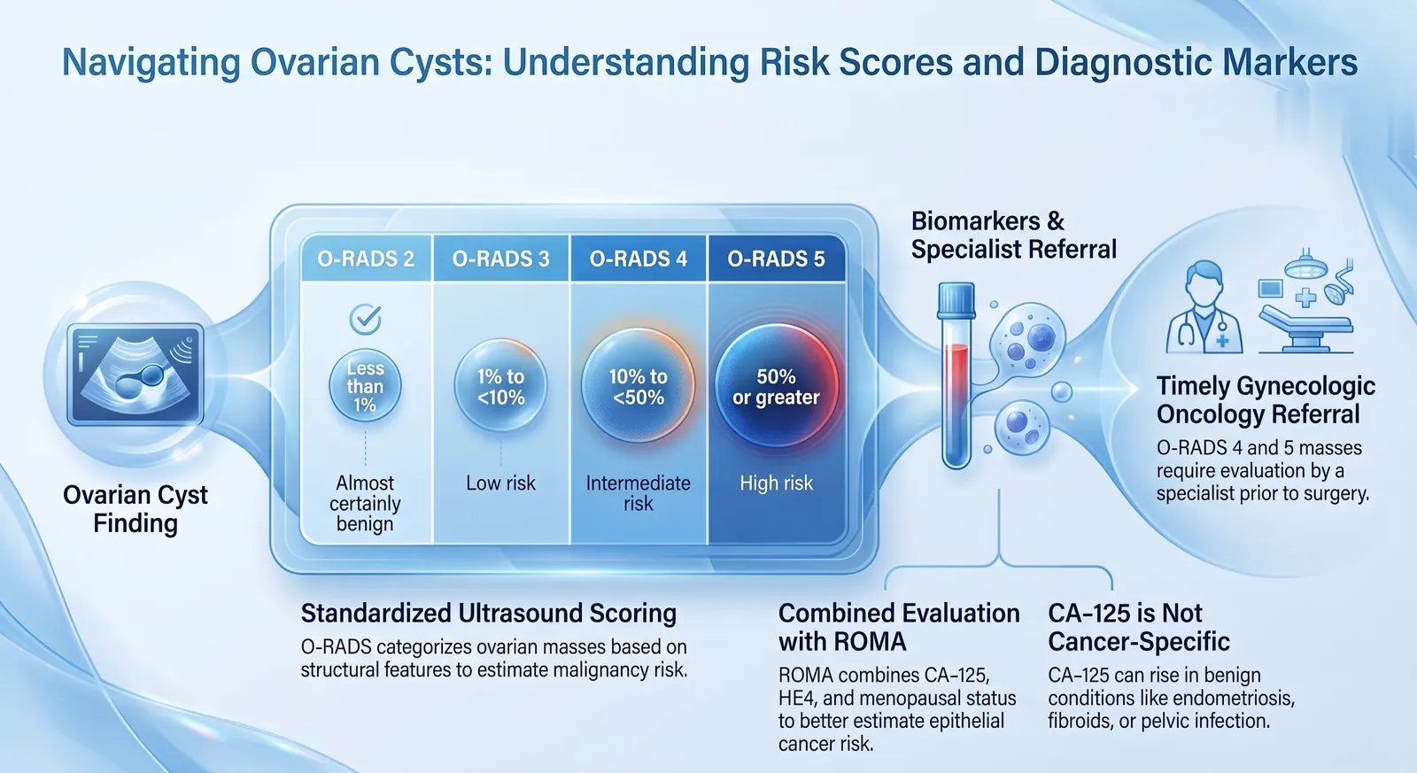
The infographic labels are in English. The percentages are the share of lesions in each O-RADS category that turn out to be cancer, not a prediction for any one person.
A scan report that says "complex cyst", a letter that says "O-RADS 4", or a CA-125 above the normal range is frightening to read. The reassuring fact is that most ovarian cysts are not cancer. The careful fact is that a few are, which is why doctors use a structured way of scoring the scan and, when needed, blood tests, MRI and sometimes surgery to be sure. This guide explains that process in plain language, and also answers the common worry about fibroids and cancer, because the two questions often arrive together.
Sudden severe pain in the lower abdomen, especially with vomiting, fainting or fever, needs urgent medical care, because a cyst can twist or burst. Bloating, early fullness or pelvic discomfort that is new and lasts more than two to three weeks also needs to be checked, whatever an earlier scan showed.
How do doctors tell a benign cyst from cancer?
They start with a transvaginal ultrasound, score what it shows using O-RADS, and then add your age and menopausal status, your symptoms, blood tests such as CA-125 and HE4 and, when the scan is unclear, an MRI. Ultrasound estimates the risk of cancer. It cannot confirm or exclude it on its own.[3]
Transvaginal scanning gives the clearest view of the ovaries. A scan through the abdomen can be enough when the cyst is large or a vaginal scan is not possible.[1] These are the features the scan reader looks for:
| Feature | More reassuring | More concerning |
|---|---|---|
| Contents | Clear fluid only, or a typical blood clot, endometrioma or dermoid pattern | Solid tissue that has its own blood flow |
| Wall | Thin and smooth | Thick or irregular |
| Chambers and septations | One chamber, or one thin smooth septation | Three or more chambers, thick or irregular septations |
| Projections | None | Papillary projections, especially four or more |
| Blood flow inside | None or minimal | Moderate to strong |
| Fluid in the abdomen | None | Ascites or nodules on the peritoneum, with no other explanation |
When features are mixed, the scoring rules tell the reader to use the more worrying feature, so that cancers are not missed.[1] That is why a "higher" score can still turn out to be benign.
What does O-RADS mean, and what do O-RADS 2, 3, 4 and 5 mean?
O-RADS is a scoring system from the American College of Radiology that turns ultrasound features into a category from 0 to 5, each with an estimated risk of cancer. O-RADS 2 is almost certainly benign (under 1%), 3 is low risk (1% to under 10%), 4 is intermediate (10% to under 50%) and 5 is high risk (50% or more).[1]
| O-RADS | Meaning | Risk of cancer | Example next step |
|---|---|---|---|
| 0 | Scan could not be assessed properly | Not applicable | Repeat the scan, or MRI |
| 1 | Normal ovary, including normal monthly follicles | 0% | Nothing needed |
| 2 | Almost certainly benign | Under 1% | Often none. A follow-up scan at 12 months for simple cysts above 5 cm before menopause or above 3 cm after menopause. A gynaecologist for symptoms |
| 3 | Low risk | 1% to under 10% | Removal if large or causing symptoms. If not removed, a scan within 6 months, then at 12 and 24 months. MRI may be added for solid lesions. A gynaecologist can manage it |
| 4 | Intermediate risk | 10% to under 50% | Specialist assessment, often with MRI, by a gynaecologist working with a gynaecological oncologist, or referral to one |
| 5 | High risk | 50% or more | Referral to a gynaecological oncologist, with further imaging (MRI and/or CT) planned by that team |
These are recommendations, not rules. The follow-up intervals above are examples of guideline-based management, not instructions for every lesion: the advice varies with the type and size of the lesion and your own situation, and the reporting radiologist and your treating doctor decide it. For example, UK RCOG guidance advises a repeat scan with CA-125 at 4 to 6 months for a simple cyst of 3 to 5 cm after menopause, where O-RADS suggests 12 months.[14] The scan category can be adjusted for symptoms, a family history or genetic risk, age, other results and the judgement of your doctors.[1] Outcomes in ovarian cancer are better when the first operation is done by a gynaecological oncologist, which is why categories 4 and 5 lead to that referral before surgery.[1]
What does O-RADS 4 mean?
An intermediate-risk lesion: roughly 1 in 10 up to 1 in 2 lesions in this group are cancer, so it is more likely to be benign than not, but too uncertain to watch. It calls for review by someone experienced, often with an MRI, and early involvement of a gynaecological oncologist.[1]
What does O-RADS 5 mean, and does it definitely mean cancer?
It is the highest-risk group, with a risk of 50% or more. It does not mean cancer is certain: some lesions in this category are benign, and only tissue examination confirms the diagnosis. It does mean you should be seen by a gynaecological oncologist promptly. Fluid in the abdomen that has no other cause, together with a category 3 to 5 lesion, also moves a score up to 5.[1]
"A higher O-RADS number means a higher chance that surgery by a specialist is the right plan. It is a reason for expert review, not a diagnosis."
Can a cyst disappear by itself, and when is surgery needed?
Yes, many cysts disappear on their own, especially functional cysts in women who still have periods.[11] There is no single size that decides surgery. Doctors weigh the scan features, symptoms, whether the cyst is growing, your age and menopausal status, and blood test results.
For cysts that are being watched, O-RADS uses a simple rule: if the cyst shrinks by about 10% to 15% or more at the first follow-up, it is considered benign and no more scans are needed. If it is stable, one more scan completes two years of stability. If it grows, scans continue and, if it keeps growing, a gynaecologist decides on treatment.[1]
Surgery is usually considered when a cyst causes pain or pressure, is very large, has suspicious features (O-RADS 4 or 5), keeps growing, or causes an emergency such as twisting or rupture.[1] When cancer is suspected, the aim is to remove the ovary with the mass intact rather than puncture it, and to avoid draining or breaking it open.[3]
Is a cyst after menopause dangerous?
Small simple cysts are common after menopause and carry a very low risk. They are followed with a scan if they are larger than 3 cm.[1] A cyst that looks complex or solid, a suspected endometrioma that appears for the first time after menopause, or a cyst with a rising CA-125 deserve closer attention, because a pelvic mass in a postmenopausal woman carries a higher chance of cancer than in a younger woman.[1][2]
What are CA-125, HE4 and ROMA?
CA-125 is a protein measured in the blood. It is raised in about 80% of advanced epithelial ovarian cancers but in only about 50% of stage I cases, so a normal result does not rule cancer out.[2] It also rises in many non-cancer conditions, so a high result does not prove cancer. HE4 is a second protein, and ROMA is a score that combines CA-125, HE4 and menopausal status to estimate whether a mass is likely to be an epithelial ovarian cancer.[6]
Can CA-125 be high without cancer?
Yes, often. Menstruation, pregnancy, endometriosis, fibroids, pelvic infection, benign ovarian cysts, liver disease and tuberculosis can all raise it.[10] This is why CA-125 is much less reliable before menopause. A usual upper limit of normal is 35 U/mL, though laboratories differ.
Can CA-125 be normal despite ovarian cancer?
Yes. About half of stage I ovarian cancers have a normal CA-125, and some types of ovarian cancer do not raise it much.[2] A normal CA-125 is not reassurance if the scan looks suspicious or symptoms continue.
What CA-125 level suggests ovarian cancer?
There is no single level that diagnoses cancer. A raised CA-125 can occur with benign conditions or with cancer, and what it means depends on how high it is, menopausal status, symptoms and the scan. A markedly raised value in a postmenopausal woman with a suspicious mass raises more concern, and a normal result does not exclude ovarian cancer.[2][14] Very high values can also occur with peritoneal tuberculosis, so the number is read together with the scan.[7]
Do I need a CA-125 for every cyst?
No. Simple, typical-looking cysts in women who still have periods generally do not need it. It is most useful in postmenopausal women with a mass, and it is not a screening test for women without symptoms.[2]
What are HE4 and ROMA used for?
HE4 is a protein that is less often raised by common non-cancer gynaecological conditions than CA-125, but it has its own limits: levels can be raised by reduced kidney function and rise with age. Neither marker is used alone. ROMA combines the two with menopausal status into one score, with separate cut-offs before and after menopause. It is meant for women who already have a pelvic mass and are being assessed for surgery, not for screening, and it must be read with the scan.[6] RCOG guidance on postmenopausal ovarian cysts does not currently recommend HE4 or ROMA for routine use, so how often they are used varies between countries and laboratories.[14] The cut-offs depend on the laboratory method, so the lab report's own interpretation matters. In women under 40 with a suspected ovarian mass, NICE advises AFP and hCG alongside CA-125 to pick out tumours that are not the common epithelial type, and LDH is sometimes added.[3][15]
Do I need an MRI, CT or PET-CT?
Not for every cyst. MRI is used when the ultrasound result is unclear, especially for solid lesions and cysts with solid parts, and it has its own O-RADS MRI score. CT is used to stage the disease once cancer is suspected. PET-CT is reserved for selected situations.[3][4]
- MRI: gives higher specificity than ultrasound for indeterminate masses, and can help avoid unnecessary surgery.[1][3]
- CT of the abdomen and pelvis (often with the chest): shows how far a suspected cancer has spread and whether surgery can remove it, and helps decide whether chemotherapy should come first.[3][4]
- PET-CT: helpful in selected cases, for example widespread peritoneal disease or raised tumour markers with inconclusive CT. It is not a routine test for a cyst.[3]
Is ultrasound enough? Is a biopsy or FNAC needed?
Ultrasound is not enough to diagnose ovarian cancer; tissue examination is needed to confirm it. When cancer is suspected in an operable mass, the usual route is surgery, which removes the mass whole, confirms the diagnosis and stages the disease at once. A needle biopsy or FNAC of the mass is generally avoided because puncturing it can spill cells and upstage the tumour.[3][14] The plan depends on how operable the mass looks, how far any disease has spread and your specialist team's assessment; in advanced disease, an image-guided core biopsy may come first.[5]
Needle sampling does have a place when the disease is already widespread or when chemotherapy is planned before surgery. Then a core biopsy is preferred, because cytology alone may not separate a borderline tumour from an invasive cancer and may not give enough material for genetic testing.[5] Fluid from the abdomen can show cancer cells, but the yield varies, roughly 30% to 70%, and fluid cytology on its own is considered acceptable only in exceptional cases when a biopsy is not possible.[3][5]
What is ascites, and can ovarian cancer be mistaken for TB or a benign cyst?
Ascites is a build-up of fluid inside the abdomen that causes swelling. It is not always cancer: liver cirrhosis, heart or kidney problems, infection and tuberculosis can cause it too. With a pelvic mass and no other explanation, it raises suspicion of ovarian cancer.[1]
In India, abdominal and peritoneal tuberculosis can closely mimic advanced ovarian cancer, with ascites, thickening of the omentum, a pelvic mass and a raised CA-125, sometimes in the hundreds. The diagnosis is usually made by a biopsy or laparoscopy, and the treatment is completely different, so tissue confirmation matters.[7][10]
The reverse also happens. Some ovarian cancers, including those that arise in endometriosis, can look like ordinary cysts on a scan.[1] That is why the report details, follow-up scans, and a second opinion from a gynaecological oncologist when something does not fit, are worth taking seriously.
Fibroid or cancer? Questions about the uterus
A fibroid is not cancer. Fibroids (leiomyomas) are very common benign muscle growths in the uterus. A rare cancer of the uterine muscle, leiomyosarcoma, can look like a fibroid on a scan, and that is the source of the worry. Leiomyosarcoma is very rare, and it is found more often in older women.[8]
Can a fibroid become cancer?
Fibroids are benign and almost all stay that way. The rare cancer that can mimic a fibroid is treated as a separate tumour.[8] Having fibroids is not a reason to expect cancer.
How can I tell a fibroid from uterine cancer?
Not reliably by ultrasound. No scan finding separates an ordinary fibroid from a leiomyosarcoma with certainty, and MRI is the best imaging tool but is also not definitive. The final answer comes from examining the removed tissue.[12] Features that make doctors look harder are rapid growth, a new fibroid-like mass after menopause, bleeding after menopause, persistent pain and unexplained weight loss.[13] Postmenopausal bleeding has its own evaluation, described in our guide to the thickened endometrium and biopsy.
Does a bulky or large uterus mean cancer?
No. "Bulky" or "enlarged" describes size, not a diagnosis. Fibroids and adenomyosis are the common reasons. It deserves a proper assessment when it comes with abnormal bleeding, pain, rapid growth or an abnormal appearance on MRI.
Can a rapidly growing fibroid be cancer?
Rarely. In a study of 1,332 women operated on for presumed fibroids, one had a leiomyosarcoma, and none of the 198 women who met a formal definition of rapid growth had a sarcoma, so growth alone is not a reliable predictor.[9] Even so, a rapidly enlarging uterine mass, particularly after menopause, deserves clinical assessment. MRI may help when the history, examination or first scan raises concern, but growth alone does not establish malignancy.[12]
Does a postmenopausal fibroid mean cancer?
No. Fibroids can be present after menopause. A fibroid that grows, or a new fibroid-like lump that appears after menopause, deserves evaluation because sarcomas are more often found in older women.[8]
Can ultrasound distinguish a fibroid from cancer?
Ultrasound can show a fibroid and may flag a suspicious pattern, such as a large solitary mass with strong blood flow, but no ultrasound finding reliably separates the two.[12] If surgery is planned for fibroids, it is reasonable to ask how the uterus will be removed, because cutting it into pieces inside the abdomen is discouraged when an unsuspected sarcoma is possible.[8]
What should I do if my report mentions a cyst, O-RADS or a raised CA-125?
Do not panic, and do not ignore it. Have the actual scan report and any blood tests reviewed by a gynaecologist, and ask for a gynaecological oncologist's opinion if the category is O-RADS 4 or 5, the cyst has solid parts, or the CA-125 is clearly raised.
Bring these to your appointment:
- The full ultrasound or MRI report, with the O-RADS category if given, the size in all three directions and any previous scans for comparison
- CA-125, HE4 or ROMA results, with the date and your menstrual or menopausal status
- Your symptoms and when they began, such as bloating, early fullness, pain, urinary or bowel change
- Family history of ovarian, breast, uterine or bowel cancer, and any genetic test result
Useful questions to ask: Which O-RADS category is this and why? Do I need an MRI? Is this a cyst to watch or to remove? If surgery is advised, will the cyst be removed whole? Our free symptom diary and questions-to-ask sheet can help you prepare, and our guide on which doctor to see explains who should assess a suspicious mass. You can also read about ovarian cancer treatment in Kolkata and our plain-language guide to ovarian cancer symptoms.
Frequently Asked Questions
Is a fibroid the same as cancer?
No. A fibroid is a benign growth of the uterine muscle. A rare cancer of the same muscle, leiomyosarcoma, can look similar on a scan, which is why the two are sometimes confused.[8]
Can a fibroid become cancer?
Fibroids are benign and almost all stay that way. The rare uterine cancer that can mimic a fibroid is treated as a separate tumour.[8]
How can I tell a fibroid from uterine cancer?
Scans cannot do it reliably. MRI helps most, and the final answer comes from examining the tissue after removal. Rapid growth, new growth after menopause, postmenopausal bleeding, persistent pain and weight loss prompt a closer look.[12][13]
Does a bulky uterus mean cancer?
No. "Bulky" describes size. Fibroids and adenomyosis are the usual causes, and cancer is a rare one that is looked for when there is abnormal bleeding, pain or rapid growth.
Can a rapidly growing fibroid be cancer?
Rarely. In a study of 1,332 women operated on for presumed fibroids, none of the 198 who met a formal definition of rapid growth had a sarcoma, so growth alone is not a reliable predictor, but quick growth still merits a specialist review.[9][13]
Does a postmenopausal fibroid mean cancer?
No. Fibroids can persist after menopause. A fibroid that grows or a new fibroid-like mass after menopause should be evaluated, because sarcomas are more common in older women.[8]
How do I know whether an ovarian cyst is benign or malignant?
Doctors score the ultrasound with O-RADS, add your age, menopausal status, symptoms and blood tests such as CA-125 and HE4, use MRI when the scan is unclear, and confirm cancer only by examining tissue.[1][3]
Is a complex ovarian cyst cancer?
Not necessarily. Complex means the cyst has septations, debris, a thick wall or a solid part. Common benign conditions such as endometriomas, dermoid cysts and cysts that have bled look complex.[1]
What does O-RADS mean?
O-RADS is the American College of Radiology's scoring system that turns ultrasound features of an ovarian or adnexal lesion into a category from 0 to 5, each with an estimated risk of cancer and a suggested next step.[1]
What does O-RADS 2 mean?
Almost certainly benign, with a risk of cancer below 1%. Many need no treatment, and some simple cysts get a follow-up scan at 12 months.[1]
What does O-RADS 3 mean?
Low risk, 1% to under 10%. The lesion may be removed if large or symptomatic, or followed with ultrasound within 6 months and then at 12 and 24 months. MRI may be added for solid lesions.[1]
What does O-RADS 4 mean?
Intermediate risk, 10% to under 50%. It is more likely benign than cancer but needs expert review, often with MRI, and early involvement of a gynaecological oncologist.[1]
Does O-RADS 5 definitely mean cancer?
No. It means a high chance, not certainty, and only tissue examination confirms the diagnosis. It does mean prompt specialist care.[1]
Does a high CA-125 mean ovarian cancer?
Not by itself. Many benign conditions raise CA-125, especially before menopause. It is read together with the scan and menopausal status.[2][10]
Can CA-125 be normal despite ovarian cancer?
Yes. About half of stage I ovarian cancers have a normal CA-125, so a normal result is not reassurance when the scan or symptoms are concerning.[2]
Is every ovarian cyst cancer?
No. Most ovarian cysts are benign, and many functional cysts disappear on their own within a few menstrual cycles.[11]
What is a simple ovarian cyst?
A thin-walled, fluid-filled, single-chamber sac with no solid parts and no internal blood flow. The chance of cancer is very low.[1]
Does a large cyst mean cancer?
No. Size alone does not mean cancer. Size is one factor in O-RADS: a typical benign lesion of 10 cm or more is generally placed in category 3, and large cysts are often removed because they cause symptoms or twist.[1]
Does a solid ovarian mass mean cancer?
Not always. A smooth solid mass with shadowing can be low risk, and a solid lump beside the ovary can be a fibroid. A solid mass with an irregular outline is high risk.[1]
Can a cyst disappear by itself?
Yes, especially functional cysts in women who have periods. If a watched cyst shrinks by about 10% to 15% or more, it is considered benign and further scans are not needed.[1][11]
When does an ovarian cyst require surgery?
When it causes pain or pressure, is very large, keeps growing, has suspicious features (O-RADS 4 or 5), or causes an emergency such as twisting or rupture.[1]
Is a cyst after menopause dangerous?
Small simple cysts are common and very low risk. A complex or solid cyst, or one that is growing, needs closer attention because a pelvic mass after menopause carries a higher chance of cancer.[1][2]
Is MRI required for an ovarian mass?
Not for every cyst. MRI is advised when ultrasound is indeterminate, especially for solid lesions and cysts with solid parts, and it has its own O-RADS MRI score.[3][4]
Is CA-125 required for every ovarian cyst?
No. Typical simple cysts in women who have periods usually do not need it. It is most useful in postmenopausal women with a mass.[2]
Is ultrasound enough to diagnose ovarian cancer?
No. Ultrasound estimates risk. The diagnosis is confirmed by examining tissue, usually from the removed mass or, in advanced disease, a core biopsy.[3][5]
Is transvaginal ultrasound better?
Generally yes for the ovaries, because it gives a closer, clearer view. A scan through the abdomen can suffice when the cyst is large or a vaginal scan is not possible.[1]
What is CA-125?
A protein measured in the blood. It is raised in about 80% of advanced epithelial ovarian cancers and about 50% of stage I cases, and it also rises in many benign conditions.[2]
What CA-125 level suggests ovarian cancer?
No single level. A usual upper limit is 35 U/mL, but a raised value can occur with benign conditions or cancer, so its meaning depends on how high it is, menopausal status, symptoms and the scan. A markedly raised level in a postmenopausal woman with a suspicious mass is more concerning, and a normal level does not exclude cancer.[2][14]
What are HE4 and ROMA?
HE4 is a second blood protein. ROMA combines CA-125, HE4 and menopausal status into a score estimating whether a pelvic mass is likely to be epithelial ovarian cancer. It is for women who already have a mass, not for screening.[6]
Is a CT scan required?
When cancer is suspected, CT of the abdomen and pelvis, often with the chest, is used to stage the disease and plan surgery. It is not needed for an ordinary cyst.[3][4]
Is PET-CT required?
No, not routinely. It is used in selected situations such as widespread peritoneal disease with inconclusive CT, or recurrence.[3]
Is a biopsy required?
For a suspected early, operable cancer, surgery that removes the mass whole is usually the diagnostic step. For advanced disease or when chemotherapy comes first, an image-guided core biopsy is advised.[3][5]
Can FNAC diagnose ovarian cancer?
FNAC of an ovarian mass is generally avoided, because puncturing it can spill cells and upstage the tumour, and cytology alone may not separate borderline from invasive cancer. Core biopsy is preferred when sampling is needed.[3][5]
Can ovarian cancer be diagnosed from fluid?
Fluid from the abdomen can show cancer cells, but the yield varies, about 30% to 70%. It is generally used when a biopsy is not possible, and tissue remains the preferred proof.[3][5]
Is ascites always cancer?
No. Cirrhosis, heart or kidney problems, infection and tuberculosis are other causes. With a pelvic mass and no other explanation, it raises suspicion of ovarian cancer.[1]
Can ovarian cancer be mistaken for tuberculosis?
Yes. In India, peritoneal tuberculosis can mimic advanced ovarian cancer, with ascites, omental thickening, a pelvic mass and a raised CA-125. A biopsy or laparoscopy tells them apart.[7]
Can ovarian cancer be mistaken for a benign cyst?
Yes. Some ovarian cancers, including those arising in endometriosis, can look like ordinary cysts on a scan. This is why follow-up and expert review matter when features do not fit.[1]
Final takeaway
- Most ovarian cysts are benign, and many go away on their own.
- O-RADS 2 to 5 is a risk score from the scan. Higher means more reason for expert review, not a diagnosis.
- CA-125 can be high without cancer and normal with cancer, so it is read together with the scan and menopausal status.
- Tissue examination is needed to confirm ovarian cancer. Whether that is done by surgery or a core biopsy depends on whether the mass looks operable, how far any disease has spread and your specialist team's assessment.
- A fibroid is not cancer, but rapid growth, new growth after menopause and postmenopausal bleeding need a proper work-up.
This article is for general education and does not replace an individual consultation. Guidelines are updated over time and differ between countries.
References
- Strachowski LM, Jha P, Phillips CH et al. O-RADS US v2022: an update from the American College of Radiology's Ovarian-Adnexal Reporting and Data System US Committee. Radiology. 2023;308(3):e230685. rsna.org
- American College of Obstetricians and Gynecologists. Practice Bulletin No. 174: Evaluation and management of adnexal masses. Obstet Gynecol. 2016;128(5):e210-e226.
- Rupa et al. Imaging recommendations for diagnosis, staging, and management of ovarian and fallopian tube cancers. Indian J Med Paediatr Oncol. 2023;44(1):100-109. thieme-connect.com
- Avesani G et al. ESR Essentials: characterisation and staging of adnexal masses with MRI and CT, practice recommendations by ESUR. Eur Radiol. 2024. ncbi.nlm.nih.gov
- Neoadjuvant chemotherapy for newly diagnosed, advanced ovarian cancer: ASCO guideline update. J Clin Oncol. 2025. ascopubs.org
- Moore RG et al. A novel multiple marker bioassay utilizing HE4 and CA125 for the prediction of ovarian cancer in patients with a pelvic mass. Gynecol Oncol. 2008;108:402-8. See also Mayo Clinic Laboratories, ROMA score test information.
- Abdominal tuberculosis mimicking ovarian cancer: a diagnostic dilemma. J Obstet Gynaecol India. ncbi.nlm.nih.gov
- US Food and Drug Administration. Updated assessment of the use of laparoscopic power morcellators to treat uterine fibroids. 2017. fda.gov
- Parker WH, Fu YS, Berek JS. Uterine sarcoma in patients operated on for presumed leiomyoma and rapidly growing leiomyoma. Obstet Gynecol. 1994;83:414-8.
- Meden H, Fattahi-Meibodi A. CA 125 in benign gynecological conditions. Int J Biol Markers. 1998;13:231-7. doi.org
- Royal College of Obstetricians and Gynaecologists / BSGE. Green-top Guideline No. 62: Management of suspected ovarian masses in premenopausal women. 2011. rcog.org.uk
- Review of leiomyoma variants. AJR Am J Roentgenol. ajronline.org
- Fersini A et al. Misdiagnosis of leiomyosarcomas: case report and medico-legal issues. Gynecol Pelvic Med. amegroups.org
- Royal College of Obstetricians and Gynaecologists. The management of ovarian cysts in postmenopausal women. Green-top Guideline No. 34. 2016; minor update December 2025. rcog.org.uk
- National Institute for Health and Care Excellence. Ovarian cancer: recognition and initial management. NICE guideline CG122. 2011; updated April 2026. nice.org.uk
Related Reading

Worried about an ovarian cyst, a high CA-125 or an O-RADS 4 or 5 report?
Dr. Dipanwita Banerjee offers consultations at CNCI Kolkata for evaluation of ovarian cysts and masses, raised tumour markers and suspected gynaecological cancer.